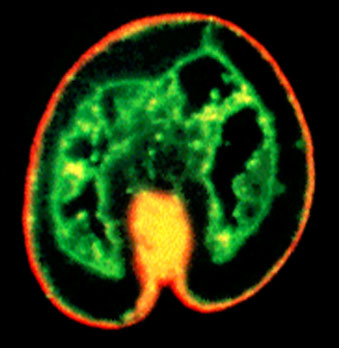
The sea urchin embryo possesses extracellular matrix layers lining the inside of the embryo (the basal lamina) and lining the outside of the embryo. The outer layer is divided into at least two layers, an apical lamina directly attached to the apical ends of the cells, and a slightly more exterior hyaline layer. In this micrograph, the basal lamina is immunostained with a monoclonal antibody (green) and the hyaline layer is immunostained with a second antibody (red).
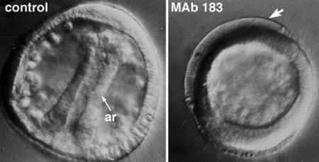
Primary invagination can be disrupted by treating embryos with a monoclonal antibody (MAb 183) that recognizes the protein hyalin, the predominant protein in the hyaline layer. The hyaline layer peels away from the epithelium in antibody treated embryos, and although mesenchyme cells can migrate in such embryos, invagination is completely blocked.
For a movie of a Mab183-treated embryo, click on the thumbnail below.
 |
|
|
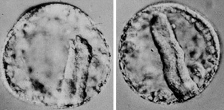
Invagination can also be disrupted by perturbing the basal lamina. Treating embryos with the lathrytic agent, b-aminoproprionitrile, results in failure of the chemical crosslinking reactions that normally occur between collagen molecules in the basal ECM. When the drug is applied during primary invagination, the result is a "kinky" archenteron with mesenchyme that have impaired motility.