The sea urchin embryo was one of the first systems in which gastrulation was studied using time-lapse cinemicrography, worked pioneered by Tryggve Gustafson and colleagues in Sweden. The simple organization of the sea urchin gastrula, combined with its transparency, make it ideal for elucidating the basic cellular principles of gastrulation. The next few pages provide an overview of the various stages of sea urchin gastrulation, and some of the cellular mechanisms that underlie them. For a brief visual discovery exercise to identify parts of the gastrula, click here.
There are several basic phases of sea urchin gastrulation. these include (1) ingression of primary, or skeletogenic, mesenchyme; (2) invagination of the vegetal plate to produce the early archenteron; (3) elongation of the archenteron, coincident with the appearance of secondary mesenchyme cells; and (4) contact of the tip of the archenteron with the animal pole region, near a thickened region of the ectoderm sometimes called the apical plate. These basic phases of gastrulation are shown diagrammatically below.
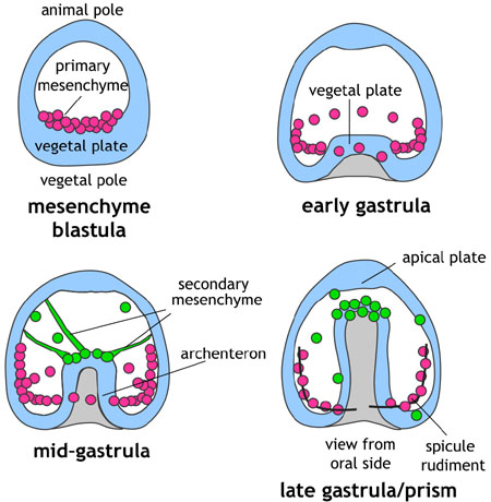
Stages of gastrulation depicted schematically. Image by Jeff Hardin, Univ. of Wisconsin.
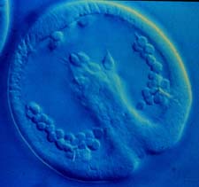
Although diagrams are helpful, actual embryos are better! Below are images of Lytechnius variegatus embryos, from mesenchyme blastula to midgastrula, courtesy of David McClay, Duke Univ.
Stages of gastrulation in Lytechinus variegatus. A. Early mesenchyme blastula. B. Skeletogenic, or primary mesenchyme (PMCs) ingressing. C. Late ingression. D. Early primary invagination of the archenteron. E. Late primary invagination. F. Onset of archenteron elongation (secondary invagination). (courtesy of David McClay, Duke Univ.)