
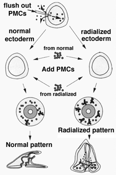
Diagram showing how PMCs can be removed and transplanted between normal and nickel-treated embryos.
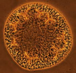
By disrupting the pattern of primary mesenchyme cells (PMCs), we can study where the positional cues reside in the embryo that generate the precise pattern PMCs normally adopt. Here nickel treatment has resulted in a radialized pattern. We can perform transplants between normal and radialized embryos to study where the cues reside that normally provide pattern information to PMCs. f the ectodermal environment provides local cues for PMC patterning, then one hypothesis regarding how nickel works is that it affects the ectoderm. If the ectoderm provides the cues that PMCs use to adopt their normal pattern, then it is possible that the ectoderm in nickel-treated embryos fails to provide the appropriate signals to the PMCs, resulting in the radialized pattern. If the predominant problem were with the ectoderm, then transplanting PMCs from a radialized embryo into normal ectoderm would be expected to yield a normally patterned skeleton. We can do this sort of experiment by flushing PMCs out of the blastocoel to create an ectodermal "shell" into which we transplant fluorescently labeled PMCs. We can place PMCs from a nickel-treated embryo into a normal shell, normal PMCs into a nickel-treated shell, etc.
As you can see in the image
below, the results of reciprocal PMC transplants are quite
clear: the ectoderm seems to provide much of the basic
information used by primary mesenchyme as they form
patterned skeletal elements. We know this because PMCs from
nickel treated embryos are able to adopt a normal pattern
when placed into a normal ectodermal shell. This is a nice
demonstration of the general principle that local
patterning cues in the environment are used by freely
migrating cells as they move, and eventually stop in
specific locations in the early embryo.
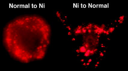
Left: fluorescently labeled PMCs from a normal embryo were placed into the ectodermal shell of an embryo treated with nickel (II) chloride; right: fluorescently labeled PMCs from a nickel-treated embryo were placed into the ectodermal shell of a normal embryo.