Multiple Wnt Signaling Pathways Converge to Orient the Mitotic Spindle in Early C. elegans Embryos
Timothy Walston,1 Christina Tuskey,2 Lois Edgar,4 Nancy Hawkins,5 Gregory Ellis,6 Bruce Bowerman,6 William Wood,4 and Jeff Hardin1,2,3
1Laboratory of Genetics, 2Program in Cellular and Molecular Biology, 3Department of Zoology, University of Wisconsin-Madison Madison, Wisconsin 53706, 4Department of Molecular, Cellular, and Developmental Biology University of Colorado-Boulder, Boulder, Colorado 80309, 5Department of Molecular Biology and Biochemistry, Simon Fraser University Burnaby, British Columbia V5A 1S6 Canada, 6Institute of Molecular Biology, University of Oregon, Eugene, Oregon 97403
Supplemental material for the following paper:
Walston, T, Tuskey, C., Edgar, E., Hawkins, N., Ellis, G., Bowerman, B., Wood, W., and Hardin, J. (2004). Multiple Wnt signaling pathways converge to orient the mitotic spindle in early C. elegans embryos. Dev Cell 7, 831-841. PubMed
Summary
Results
Video sequence 1: wild-type and mutant ABar division - tubulin::gfp
Additional footage not included with published supplementary materials (courtesy of Tim Walston):
Video sequence 2: wild-type ABar division (DIC)
Video sequence 3: mutant ABar division (DIC)
Video sequence 4: wild-type ABar division - contact by C
Summary
How cells integrate the input of multiple polarizing signals during division is poorly understood. We demonstrate that two distinct Caenorhabditis elegans Wnt pathways contribute to the polarization of the ABar blastomere by differentially regulating its duplicated centrosomes. Contact with the C blastomere orients the ABar spindle through a non-transcriptional Wnt spindle alignment pathway, while a Wnt/b-catenin pathway controls the timing of ABar spindle rotation. The three C. elegans Dishevelled homologs contribute to these processes in different ways, suggesting that functional distinctions may exist among them. We also find that CKI (KIN-19) not only plays a role in the Wnt/b-catenin pathway, but in the Wnt spindle orientation pathway as well. Based on these findings, we establish a model for the coordination of cell-cell interactions and distinct Wnt signaling pathways that ensures the robust timing and orientation of spindle rotation during a developmentally regulated cell division event.
Return to top
Results
Introduction to the video sequences
During development, certain cell divisions must occur with a specific orientation to form complex structures and body plans. In many cases, the polarizing input for oriented divisions involves Wnt signaling. The orientation of blastomere divisions in the early C. elegans embryo has been shown to require Wnt signaling. In the 4-cell embryo, the EMS blastomere is induced by its posterior neighbor, the P2 blastomere. This induction has two consequences: it specifies the fates of EMS daughter cells (E and MS) and properly positions the mitotic spindle of EMS. The fates of MS and E are controlled in part by a Wnt signaling pathway that regulates the activity of the Tcf/Lef transcription factor, POP-1, in conjunction with the b-catenin, WRM-1. The orientation of the EMS division is controlled by a transcription-independent Wnt pathway involving a Wnt (MOM-2), Porcupine (Porc; MOM-1), Fz (MOM-5), and GSK-3, the C. elegans GSK-3b homolog. A pathway involving MES-1, a receptor tyrosine kinase, and SRC-1, a Src family tyrosine kinase, acts redundantly with Wnt signaling with respect to the fate of EMS daughters and the orientation of the EMS spindle.
In addition to regulating the orientation of the EMS division, loss of function of four of the mom (more mesoderm) genes, mom-1 (Porc), mom-2 (Wnt), mom-5 (Fz), and mom-3 (uncloned) cause spindle alignment defects in the ABar blastomere of the 8-cell embryo. In this study, we demonstrate the role of two Wnt signaling pathways involved in regulating the ABar mitotic spindle. First, the non-transcriptional Wnt spindle alignment pathway requires contact from the C blastomere to align the spindle of ABar. We show that the three Dshs differentially participate in aligning the spindles of EMS and ABar, and vary with respect to their interaction with the Src signaling pathway during spindle orientation. Second, a Wnt/b-catenin pathway regulates the timing of spindle rotation in ABar, presumably by specifying the fate of neighboring blastomeres. Taken together, these studies indicate that spindle orientation during early development is a tightly regulated event, influenced by multiple cues transmitted via redundant pathways.
Return to top
Video sequence 1:
Spindle misalignment and delayed rotation
phenotypes in the ABar blastomere
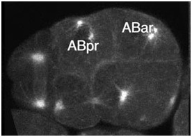
Confocal movie of ABar spindle alignment visualized using TBB-2/b-tubulin::GFP. In wild-type embryos (left), the ABar spindle rotates quickly into the proper position perpendicular to that of ABpr, while wrm-1(RNAi) embryos (middle) display a delay in spindle rotation. The ABar spindle in dsh-2(RNAi); mig-5(RNAi) embryos (right) fails to rotate into the proper position. All embryos are shown in right-hand views with anterior to the right and dorsal at the top. Time points are 1 minute apart. Scale bar = 10 um.
Click on the link to load video sequence 1: Video sequence #1 (1.2 Mb)
Return to top
Video sequence 2:
ABar orientation in a wild-type embryo
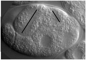
Nomarski movie of ABar spindle alignment (right-hand view; anterior to the right and dorsal at the top; movie courtesy of Tim Walston). In wild-type embryos, the ABar spindle rotates quickly into the proper position perpendicular to that of ABpr (spindle orientations are shown as black lines).
Click on the link to load video sequence 2: Video sequence #2 (7.8 Mb)
Return to top
Video sequence 3:
ABar misalignment in a Dsh mutant embryo
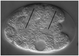
Nomarski movie of ABar spindle misalignment in a dsh-2(or302);mig-5(RNAi);dsh-1(RNAi) embryo (right-hand view; anterior to the right and dorsal at the top; movie courtesy of Tim Walston). The ABar spindle in fails to rotate into the proper position (spindle orientations are shown as black lines).
Click on the link to load video sequence 3: Video sequence #3 (10.2 Mb)
Return to top
Video sequence 4:
ABar alignment is induced by contact with C
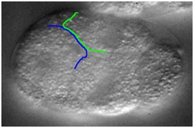
Two different focal planes from a 4d Nomarski movie show the orientation of ABar (right) relative to time of contact with C (left). In wild-type embryos, the ABar spindle rotates quickly into the proper position perpendicular to that of ABpr (spindle orientation is shown as black lines). This coincides with contact by C; the edges of C (blue) and ABar (green) are shown.
Click on the link to load video sequence 4: Video sequence #4 (4.1 Mb)
Timothy Walston,1 Christina Tuskey,2 Lois Edgar,4 Nancy Hawkins,5 Gregory Ellis,6 Bruce Bowerman,6 William Wood,4 and Jeff Hardin1,2,3
1Laboratory of Genetics, 2Program in Cellular and Molecular Biology, 3Department of Zoology, University of Wisconsin-Madison Madison, Wisconsin 53706, 4Department of Molecular, Cellular, and Developmental Biology University of Colorado-Boulder, Boulder, Colorado 80309, 5Department of Molecular Biology and Biochemistry, Simon Fraser University Burnaby, British Columbia V5A 1S6 Canada, 6Institute of Molecular Biology, University of Oregon, Eugene, Oregon 97403
Supplemental material for the following paper:
Walston, T, Tuskey, C., Edgar, E., Hawkins, N., Ellis, G., Bowerman, B., Wood, W., and Hardin, J. (2004). Multiple Wnt signaling pathways converge to orient the mitotic spindle in early C. elegans embryos. Dev Cell 7, 831-841. PubMed
Summary
Results
Video sequence 1: wild-type and mutant ABar division - tubulin::gfp
Additional footage not included with published supplementary materials (courtesy of Tim Walston):
Video sequence 2: wild-type ABar division (DIC)
Video sequence 3: mutant ABar division (DIC)
Video sequence 4: wild-type ABar division - contact by C
Summary
How cells integrate the input of multiple polarizing signals during division is poorly understood. We demonstrate that two distinct Caenorhabditis elegans Wnt pathways contribute to the polarization of the ABar blastomere by differentially regulating its duplicated centrosomes. Contact with the C blastomere orients the ABar spindle through a non-transcriptional Wnt spindle alignment pathway, while a Wnt/b-catenin pathway controls the timing of ABar spindle rotation. The three C. elegans Dishevelled homologs contribute to these processes in different ways, suggesting that functional distinctions may exist among them. We also find that CKI (KIN-19) not only plays a role in the Wnt/b-catenin pathway, but in the Wnt spindle orientation pathway as well. Based on these findings, we establish a model for the coordination of cell-cell interactions and distinct Wnt signaling pathways that ensures the robust timing and orientation of spindle rotation during a developmentally regulated cell division event.
Return to top
Results
Introduction to the video sequences
During development, certain cell divisions must occur with a specific orientation to form complex structures and body plans. In many cases, the polarizing input for oriented divisions involves Wnt signaling. The orientation of blastomere divisions in the early C. elegans embryo has been shown to require Wnt signaling. In the 4-cell embryo, the EMS blastomere is induced by its posterior neighbor, the P2 blastomere. This induction has two consequences: it specifies the fates of EMS daughter cells (E and MS) and properly positions the mitotic spindle of EMS. The fates of MS and E are controlled in part by a Wnt signaling pathway that regulates the activity of the Tcf/Lef transcription factor, POP-1, in conjunction with the b-catenin, WRM-1. The orientation of the EMS division is controlled by a transcription-independent Wnt pathway involving a Wnt (MOM-2), Porcupine (Porc; MOM-1), Fz (MOM-5), and GSK-3, the C. elegans GSK-3b homolog. A pathway involving MES-1, a receptor tyrosine kinase, and SRC-1, a Src family tyrosine kinase, acts redundantly with Wnt signaling with respect to the fate of EMS daughters and the orientation of the EMS spindle.
In addition to regulating the orientation of the EMS division, loss of function of four of the mom (more mesoderm) genes, mom-1 (Porc), mom-2 (Wnt), mom-5 (Fz), and mom-3 (uncloned) cause spindle alignment defects in the ABar blastomere of the 8-cell embryo. In this study, we demonstrate the role of two Wnt signaling pathways involved in regulating the ABar mitotic spindle. First, the non-transcriptional Wnt spindle alignment pathway requires contact from the C blastomere to align the spindle of ABar. We show that the three Dshs differentially participate in aligning the spindles of EMS and ABar, and vary with respect to their interaction with the Src signaling pathway during spindle orientation. Second, a Wnt/b-catenin pathway regulates the timing of spindle rotation in ABar, presumably by specifying the fate of neighboring blastomeres. Taken together, these studies indicate that spindle orientation during early development is a tightly regulated event, influenced by multiple cues transmitted via redundant pathways.
Return to top
Video sequence 1:
Spindle misalignment and delayed rotation
phenotypes in the ABar blastomere
Confocal movie of ABar spindle alignment visualized using TBB-2/b-tubulin::GFP. In wild-type embryos (left), the ABar spindle rotates quickly into the proper position perpendicular to that of ABpr, while wrm-1(RNAi) embryos (middle) display a delay in spindle rotation. The ABar spindle in dsh-2(RNAi); mig-5(RNAi) embryos (right) fails to rotate into the proper position. All embryos are shown in right-hand views with anterior to the right and dorsal at the top. Time points are 1 minute apart. Scale bar = 10 um.
Click on the link to load video sequence 1: Video sequence #1 (1.2 Mb)
Return to top
Video sequence 2:
ABar orientation in a wild-type embryo
Nomarski movie of ABar spindle alignment (right-hand view; anterior to the right and dorsal at the top; movie courtesy of Tim Walston). In wild-type embryos, the ABar spindle rotates quickly into the proper position perpendicular to that of ABpr (spindle orientations are shown as black lines).
Click on the link to load video sequence 2: Video sequence #2 (7.8 Mb)
Return to top
Video sequence 3:
ABar misalignment in a Dsh mutant embryo
Nomarski movie of ABar spindle misalignment in a dsh-2(or302);mig-5(RNAi);dsh-1(RNAi) embryo (right-hand view; anterior to the right and dorsal at the top; movie courtesy of Tim Walston). The ABar spindle in fails to rotate into the proper position (spindle orientations are shown as black lines).
Click on the link to load video sequence 3: Video sequence #3 (10.2 Mb)
Return to top
Video sequence 4:
ABar alignment is induced by contact with C
Two different focal planes from a 4d Nomarski movie show the orientation of ABar (right) relative to time of contact with C (left). In wild-type embryos, the ABar spindle rotates quickly into the proper position perpendicular to that of ABpr (spindle orientation is shown as black lines). This coincides with contact by C; the edges of C (blue) and ABar (green) are shown.
Click on the link to load video sequence 4: Video sequence #4 (4.1 Mb)