 THE HARDIN LAB
The C. elegans Zonula Occludens Ortholog Cooperates with the Cadherin Complex to Recruit Actin during Morphogenesis
Christina Lockwood1,3, Ronen Zaidel-Bar2, and Jeff Hardin1,2,4
1Program in Cellular and Molecular Biology, and 2Department of Zoology, University of Wisconsin Madison, WI 53706
3Present address: Washington University School of Medicine, Department of Pathology & Immunology, 660 S Euclid, Campus Box 8118 St. Louis, MO 63110
4Corresponding author
Supplemental material for the following paper:
Lockwood, C., Zaidel-Bar, and Hardin, J. (2008). The C. elegans Zonula Occludens Ortholog cooperates with the cadherin complex to recruit actin during morphogenesis. Curr. Biol.18:1333-7. PubMed
Summary
Supplemental Figure 1
Supplemental Figure 2
Supplemental Figure 3
Supplemental Figure 4
Supplemental Figure 5
Movie 1
Movie 2
Movie 3
Movie 4
Movie 5
Movie 6
Movie 7
Movie 8
Movie 9
Movie 10
Summary
The dramatic cell shape changes necessary to form a multicellular organism require cell-cell junctions to be both pliable and strong. The Zonula Occludens (ZO) subfamily of membrane-associated guanylate kinases (MAGUKs) are scaffolding molecules thought to regulate cell-cell adhesion [1-3], but there is little known about their roles in vivo. To elucidate the functional role of ZO proteins in a living embryo, we have characterized the sole C. elegans ZO family member, ZOO-1. ZOO-1 localizes with the cadherin-catenin complex during development and its junctional recruitment requires the transmembrane proteins HMR-1/E-cadherin and VAB-9/claudin, but surprisingly, not HMP-1/α-catenin or HMP-2/β-catenin. zoo-1 knockdown results in lethality during elongation, resulting in the rupture of epidermal cell-cell junctions under stress and failure of epidermal sheet sealing at the ventral midline. Consistent with a role in recruiting actin to the junction in parallel to the cadherin-catenin complex, zoo-1 loss of function reduces the dynamic recruitment of actin to junctions and enhances the severity of actin filament defects in hypomorphic alleles of hmp-1 and hmp-2. These results show that ZOO-1 cooperates with the cadherin-catenin complex to dynamically regulate strong junctional anchorage to the actin cytoskeleton during morphogenesis.
Supplemental Figures
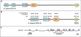
Supplementary Figure 1. zoo-1 encodes a C. elegans Zonula Occludens Ortholog.
Although the overall amino acid similarity of ZOO-1A to Homo sapiens ZO-1A is 34.2%, individual domains show much higher conservation. (A) Schematic structure of H. sapiens ZO-1A, which contains the following domains: PDZ, PSD-95, Dlg-1, ZO-1; SH3, Src Homology 3; GuK, Guanylate Kinase; ZU5, ZO-1, UNC-5 proline-rich region. (B) Schematic structure of ZOO-1. Amino acid residues of ZOO-1A corresponding to each region based on SMART predictions are indicated, as is the percent amino acid similarity within each domain compared to H. sapiens ZO-1A. (C) Structure of the zoo-1 gene is shown with exons represented by boxes. Alleles gk404 and cxTi8317 are indicated.
Supplemental Figure 1 - JPEG (960 Kb) Download
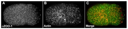
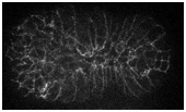
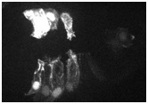
Supplemental Figure 2. ZOO-1 colocalizes with actin at cell borders during morphogenesis.
Confocal images of embryos stained for ZOO-1 (A,D,G), F-actin (B,E,H), and the merge (C,F). (A-C) Embryo beginning ventral enclosure. (D-F) Embryo at the 1.25-stage of elongation has increased ZOO-1 at cell borders. (G, H) Embryo at the 2-fold stage of elongation has robust junctional ZOO-1. Insets in G and H are enlargements of the boxed regions. Scale bar, 10 μm.
Supplemental Figure 2 - JPEG (430 Kb) Download
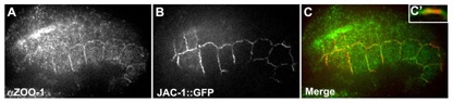
Supplemental Figure 3. ZOO-1 colocalizes with the cadherin complex in the epidermis.
(A-C) Confocal images showing a comma stage embryo stained for ZOO-1 (A) that is also expressing JAC-1/p120-catenin::GFP (B). The merged image is shown in C (ZOO-1, red; JAC-1, green). C' shows a projected region of a junction in sagittal profile enlarged 3X. (D-F) Confocal images of a 1.5-fold stage embryo stained for ZOO-1 (D) and AJM-1 (E). The merged image is shown in F (ZOO-1, red; AJM-1, green). F' shows a projected region of a junction in sagittal profile enlarged 3X. Scale bar, 10 μm. Quantitative colocalization analysis confirms that ZOO-1 colocalizes with the cadherin complex. For JAC-1, r2 = 0.72 ± 0.05, mean ± SEM, n = 3, whereas for AJM-1, r2 = 0.39 ± 0.05, n = 3 (significantly different from JAC-1, p < 0.004, Fisher’s exact test).
Supplemental Figure 3 - JPEG (330 Kb) Download
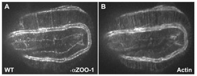
Supplemental Figure 4. zoo-1(RNAi);rrf-3(pk1426) gene knockdown abolishes antibody signal.
Embryos are stained for ZOO-1 (left column) and F-actin (right column). (A, B) Wild-type two-fold embryo corresponding to Figure 3E. (C, D) Two-fold zoo-1(RNAi) embryo, corresponding to Figure 3F. (E, F) Three-fold rrf-3(pk1426) embryo. (G, H) Three-fold zoo-1(RNAi);rrf-3(pk1426) embryo. Compare residual levels of ZOO-1 in a zoo-1(RNAi) background (C) versus complete absence of ZOO-1 in zoo-1(RNAi);rrf-3(pk1426) (G). Scale bar, 10 μm.
Supplemental Figure 4 - JPEG (480 Kb) Download
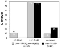
Supplemental Figure 5. Loss of zoo-1 function enhances rupture defects in mel-11(it26) hypercontractile mutants.
The percentage arrested at various stages of elongation was scored for zoo-1(RNAi); mel-11(it26) and mel-11(it26) embryos. Asterisk: significantly different, p < 0.001 (Fisher's exact test).
Supplemental Figure 5 - JPEG (230 Kb) Download
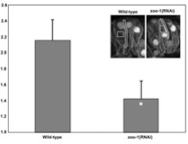
Supplemental Figure 6. zoo-1(RNAi);rrf-3 embryos accumulate less actin at cell borders.
The ratio of junctional to cytoplasmic actin was measured as described in Materials and Methods. Sample ROIs for representative wild-type and zoo-1(RNAi);rrf-3 embryos are shown (inset). Asterisk: significantly different, p < 0.01(two-tailed student t-test).
Supplemental Figure 6 - JPEG (100 Kb) Download
Return to top
Supplemental Movies
Movie 1. ZOO-1::GFP in a wild-type embryo.
Frames were collected at 200-second time intervals for 90 minutes. Movie is displayed at a rate of 7 frames per second.
(3.2 Mb) Download
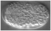
Movie 2. Elongation in a wild-type embryo.
Frames were collected at 6-minute time intervals for 4 hours. Movie is displayed at a rate of 7 frames per second.
(1 Mb) Download
Movie 3. Elongation in an rrf-3(pk1426) embryo.
Frames were collected at 6-minute time intervals for 4 hours. Movie is displayed at a rate of 7 frames per second.
(0.4 Mb) Download
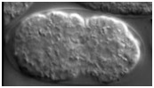
Movie 4. An elongation rupture phenotype in a zoo-1(RNAi);rrf-3(pk1426) embryo.
Frames were collected at 6-minute time intervals for 3 hours. Movie is displayed at a rate of 7 frames per second.
(0.3 Mb) Download
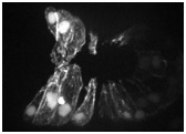
Movie 5. F-actin dynamics during ventral enclosure in a wild-type embryo expressing vab-10::ABD::GFP.
The boxed regions show extensive actin accumulation between two pairs of ventral epidermal cells (ABpr/laappap and ABpr/laappp). The pair between these two pairs (ABpr/laapppa) immediately fuses in wild-type embryos to form a syncytium. Frames were collected at 60-second time intervals. Elapsed time in seconds is shown.
(2.6 Mb) Download
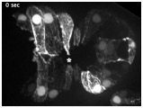
Movie 6. F-actin dynamics in a zoo-1(RNAi);rrf-3(pk1426) embryo expressing vab-10::ABD::GFP.
The asterisk show a region in which ventral midline defects occur. The boxed region shows ABpr/laappp, which have failed to make contact at the midline. Frames were collected at 60-second time intervals. Elapsed time in seconds is shown.
(0.7 Mb) Download
Movie 7. F-actin dynamics in a zoo-1(RNAi);rrf-3(pk1426) embryo expressing vab-10::ABD::GFP.
The boxed region shows ABpr/laapppa, which have failed to make contact at the midline, and hence have not fused. Frames were collected at 60-second time intervals. Elapsed time in seconds is shown.
(1.3 Mb) Download
Supplemental Movie 8. An elongation arrest phenotype in a hmp-1(fe4) embryo.
Frames were collected at 6-minute time intervals for 4 hours. Movie is displayed at a rate of 7 frames per second.
(0.4 Mb) Download
Supplemental Movie 9. A Humpback phenotype in a zoo-1(RNAi);hmp-1(fe4) embryo.
Frames were collected at 6-minute time intervals for 4 hours. Movie is displayed at a rate of 7 frames per second.
(0.3 Mb) Download
Supplemental Movie 10. An elongation rupture phenotype in a zoo-1(RNAi);hmp-(fe4) embryo.
(0.3 Mb) Download
Return to top