
 |
We study how sheets of cells (epithelial cells) rearrange, migrate, and adhere to one another in the forming epidermis, or hypodermis, of the early C. elegans embryo. We use a variety of approaches to study epithelial morphogenesis, including genetics, genomics, and advanced microscopy. Because the events we study occur in all animal embryos, what we are discovering has relevance for understanding fundamental processes during normal embryonic development. By studying what molecular processes control cell movements and cell adhesion, we hope to shed light on basic mechanisms of cancer metastasis, and on the events that lead to common birth defects. |
|
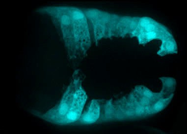
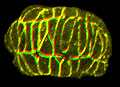
A C. elegans embryo undergoing ventral enclosure expressing a dlg-1p::gfp transcriptional reporter pseudocolored cyan, imaged using a Nikon 1.45 NA TIRF objective on our Perkin-Elmer UltraView spinning disk confocal microscope [Mark Sheffield].
|
|
We study three major events, all of which occur in virtually all animal embryos (click to learn more):
|
||
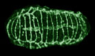 |
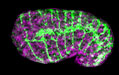
(1) Dorsal intercalation: directed cell rearrangement |
Dorsal hypodermal cells comprise two rows of epithelial cells that lie along the dorsal midline and extend along much of the anterior-posterior axis. These cells intercalate to form a single row of cells. |
 |
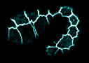
(2) Ventral enclosure: epithelial sheet sealing |
Ventral hypodermal cells migrate ventrally and meet at the ventral midline. Specific cells lead this migration, which is completed when cells make cadherin-dependent contacts along the ventral midline. |
 |
(3) Junctional maturation: organizing cell adhesion complexes |
Morphogenesis requires that cells make strong connections with one another. We are studying how two complexes, the cadherin complex and the DLG-1/AJM-1 complex are regulated during morphogenesis. |
|
We are also developing advanced microscopy techniques that have broad applications, including:
|
||
 |
(4) 4d microscopy tools | We use the plugin architecture of the public domain program, ImageJ, to create easy to use software at no cost. Click here to learn more... |
 |
(5) Correlative EM | We have developed ways to perform 4d microscopy on living embryos expressing GFP-tagged transgenes, followed by high-pressure freezing and immunogold labeling. To learn more, click here. |