 THE HARDIN LAB
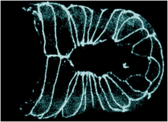 A C. elegans embryo undergoing ventral enclosure expressing DLG-1::GFP [Mathias Koeppen].
Ventral epidermal cells migrate ventrally and meet at the ventral midline. Specific cells lead this migration, which is completed when cells make cadherin-dependent contacts along the ventral midline. Click on the links to see example videos…
Ventral enclosure in a wild-type embryo visualized using Nomarski microscopy (Williams-Masson et al., 1997; PubMed). Anterior to the left. Frames were acquired at 2 min. intervals. Original movie footage courtesy of Mathias Koeppen.
Download | Play movie
Ventral enclosure in a wild-type embryo visualized using a dlg-1::gfp translational fusion (Koeppen et al., 2001; PubMed). Frames were acquired at 5 min intervals. Footage courtesy of Mathias Koeppen
Download | Play movie
We showed that ventral enclosure is driven by migration of leading edge cells (Williams-Masson et al, 1997. Development 124, 2889-2901; Sheffield et al, 2007. Curr. Biol. 17, 1791-1796), and that successful enclosure requires cell-cell adhesion as cells meet on the central midline (Raich et al, 1999. Curr Biol 9, 1139-1146). We are studying:
(1) Protrusive activity at the leading edge of migrating ventral cells;
(2) How cells create new junctional connections at the ventral midline; and
(3) How formation of cellular rosettes organizes neuroblasts over which ventral epidermal cells migrate, following up on our recent work (Serre et al, 2023. PLoS Genet. 2023 Mar 3;19(3):e1010507).
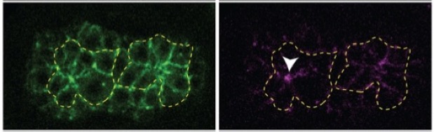 Cellular rosettes during ventral cleft closure ( SRGP-1::mNG, HMP-1::mScarlet; J. Serre)