


Cleaving cells use the same polymer systems to perform mitosis and cytokinesis as dividing somatic cells. In the micrograph shown here, kindly provided by Dr. Jon Holy, microtubules have been immunostained, and DNA has been stained using a dye. One major unsolved problem in embryonic cleavage is why the cytokinetic furrows form at particular locations, and why the spindles are oriented along particular planes of cleavage. One idea regarding the former has been suggested by the work of Dr. Ray Rappaport in sand dollars (very similar to sea urchins). His ideas have become known as the "astral stimulation" model; in this model, microtubules from the mitotic asters stimulate furrow formation.
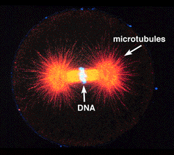
Cleaving 1-cell zygote, double labeled for DNA (light blue) and microtubules (red). Note that the astral microtubules radiate away from the spindle poles towards the cortex (astral microtubules are denoted by the arrow below the word "microtubules" in the picture).