


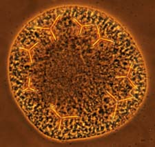
Left - A normal L. variegatus pluteus, show the bilateral skeletal elements (spicules). Right - A radialized embryos resulting from treatment with 0.5 mM nickel (II) chloride from the 2-cell to early gastrula stages.
By disrupting the pattern of primary mesenchyme cells (PMCs), we can study where the positional cues reside in the embryo that generate the precise pattern PMCs normally adopt. Here nickel treatment has resulted in a radialized pattern. As the next page shows, we can perform transplants between normal and radialized embryos to study patterning.