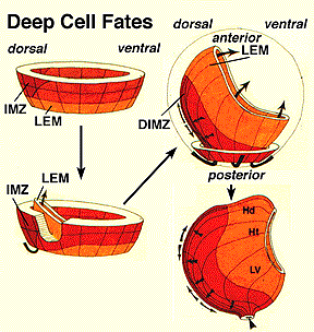


The montage below (modifed from diagrams originally made by Dr. Ray Keller, Univ. of Virginia) provides an overview of the deep cells of the marginal zone during gastrulation in Xenopus. Deep cell movements can be divided into several steps: (1) As the dorsal lip of the blastopore forms, leading edge mesoderm (shown in orange here) is rotated up into the interior of the embryo as "tongue" of material, probably as result of the apical constriction and bending of the blastopore lip by bottle cells. (2) The deep cells of the involuting marginal zone (shown in red here), then move into the interior via involution, as they undergo convergence and extension. (3) Convergence and extension is highly asymmetric; the dorsal IMZ cells (shown on the left here) converge and extend much more dramatically than the ventral and lateral regions of the IMZ. This results in preferential elongation of the embryo along the forming anterior-posterior axis. Also note that by the end of this process, the deep cells of the original marginal zone have spread to create a new middle layer of tissue within the embryo, the mesoderm. The dorsal IMZ cells produce the axial mesoderm including notochord and somites. In addition, the leading edge cells have moved all the way to the anterior region of the embryo. Dorsal leading edge cells will form structures of the head.