


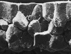
 The sequence of scanning electron micrographic images at the right
provides an overview of the behavior of the animal cap during Xenopus
gastrulation. Each image is a fracture through the roof of the
blastocoel
at various times during gastrulation. Note the thinning of the
entire tissue, which involves radially directed intercalation of deep cells
and flattening of the superficial
epithelium.
The sequence of scanning electron micrographic images at the right
provides an overview of the behavior of the animal cap during Xenopus
gastrulation. Each image is a fracture through the roof of the
blastocoel
at various times during gastrulation. Note the thinning of the
entire tissue, which involves radially directed intercalation of deep cells
and flattening of the superficial
epithelium.
Scanning electron micrographs of the animal cap during epiboly