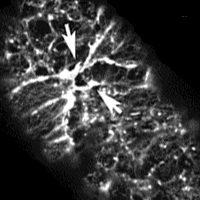
Our work with C. elegans focuses on the genetic basis of epithelial morphogenesis in the forming epidermis, or hypodermis, of the early embryo. The dorsal hypodermal cells comprise two rows of epithelial cells that lie along the dorsal midline and extend along much of the anterior-posterior axis. These cells intercalate to form a single row of cells; at the same time, the ventral hypodermal cells migrate ventrally and meet at the ventral midline. We are now studying these processes using "4-dimensional" videomicroscopy (i.e., collection of multiple focal planes at each time point, using a computer-controlled focus motor), immunofluorescence, and laser microsurgery to disable specific cells involved in these movements. With the use of this technique, we have identified specific cells that involved
in the migration of the hypodermis ventrally.
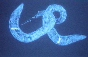
Using the phenotypes from our laser ablation experiments, we have begun an extensive screen for mutations which alter these process in specific ways. At the same time, we are characterizing cloned genes that encode proteins thought to be involved in epithelial morphogenesis in other animals. These include the protein alpha catenin, which is thought to link cadherins to the actin cytoskeleton. In collaboration with M. Costa and J. Priess (Fred Hutchinson Cancer Res. Center., Seattle), we have shown that mutations in a novel alpha catenin result in specific defects in hypodermal enclosure and subsequent elongation of the embryo into a worm-shaped larva.