

The slow block to polyspermy in the sea urchin embryo consists of a physical barrier to further sperm penetration into the egg. Cortical granule exocytosis results in the formation of the fertilization envelope (often called the fertilization "membrane", even though the structure is not a true membrane). The fertilization envelope is formed by the lifting of the vitelline envelope away from the egg plasma membrane. The cortical granules contain enzymes that aid in the detachment of the vitelline envelope, as well as other components that aid the osmotic swelling of the fertilization envelope away from the egg. Cortical granules also contain extracellular matrix proteins that are deposited on the egg surface, including the protein hyalin, which is the major component of the hyaline layer.
Cortical granule have a lamellar (layered) appearance in transmission electron micrographs.
The animation shown here simulates the fusion of the cortical granule membrane with the egg plasma membrane, resulting in the dumping of the cortical granule's contents to the exterior of the fertilized egg (courtesy of Dr. David Epel).
|
|
Animation of vesicle fusion: |
Animation of early events in fertilization, including cortical granule exocytosis: |
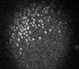
The movie shown here uses a lipophilic dye to visualize individual cortical granule fusion events (courtesy of Dr. Mark Terasaki, Univ. of Connecticut). As you can see, fusions sweep over the egg surface, beginning with the site of sperm entry.
 Quicktime movie of cortical granule release (715 K)