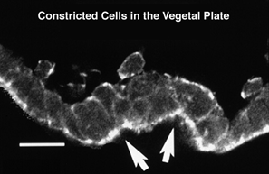
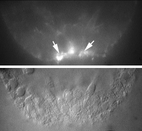
Evidence for apical constriction comes from several sources. Here the vegetal plate in L. pictus has been stained with phalloidin to reveal actin microfilaments. The arrows point to two regions of constriction that make local "kinks" in the vegetal plate. Robert Burke and colleagues at the University of Victoria were the first to show that a dense localization of actin is present in these apically constricted. The picture below shows an example of this from the species they work on, S. purpuratus. (the micrograph was generated in our laboratory).
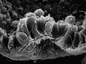
The picture below is a scanning electron micrograph of a comparable vegetal plate in L. pictus, which also shows the kinks at locations of bottle cells formation. Since these are sagittal sections through the vegetal plate, if these cells formed all the way around the vegetal plate, we would expect a ring of such cells if we could look at the surface of the vegetal plate from the bottom (i.e., the vegetal pole). The next page shows how we can do this...