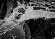


A high magnification view of L. variegatus late gastrula processed for scanning electron microscopy, showing details of a filopodium extended by a secondary mesenchyme cell (micrograph courtesy of Dr. John Morrill, Univ. of South Florida)
The onset of secondary invagination correlates with the appearance of long, thin filopodia extended by secondary mesenchyme cells at the tip of the archenteron. Under a variety of circumstances, there was substantial evidence that SMCs were required for extension of the archenteron during secondary invagination. The next several pages explore the role of secondary mesenchyme cells in completing the elongation of the archenteron...