
Sticky Worms: Adhesion Complexes in C. elegans
Elisabeth A. Cox and Jeff Hardin
Department of Zoology, University of Wisconsin, Madison, WI 53706
Address correspondence to J. Hardin, Department of Zoology, University of Wisconsin, 1117 W. Johnson St., Madison, WI . E-mail: jdhardin@wisc.edu
Supplemental material for the following paper:
Cox, E.A. and Hardin, J. (2004). Sticky worms: adhesion complexes in C. elegans. J Cell Sci. 117, 1885-1897. Medline
Summary
Video sequence 1: Protrusions during ventral enclosure
Video sequence 2: Ventral enclosure failure in a Hmr embryo
Video sequence 3: Elongation failure in a Hmp embryo
Video sequence 4: Elongation in a wild-type embryo
Summary
Caenorhabditis elegans is a powerful model system for investigating the establishment, regulation and function of adhesive structures in vivo. C. elegans has several adhesion complexes related to those in vertebrates. These include: (1) epithelial apical junctions, which have features of both adherens and tight junctions; (2) dense bodies, which are muscle-attachment structures similar to focal adhesions; (3) fibrous organelles, which resemble hemidesmosomes and mediate mechanical coupling between tissues; and (4) a putative dystrophin-glycoprotein complex that has potential roles in muscle function and embryogenesis. Recent work has increased our understanding of these structures and has given new insights into the functions of their vertebrate counterparts.
Return to top
Video sequence 1: Protrusions during ventral enclosure
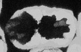 |
A projected multiphoton movie showing protrusive activity in a living embryo during ventral enclosure. The embryo is expressing an epithelial-specific cytoplasmic GFP. |
Click on the link to load video sequence 1: Video sequence #1 (0.75 Mb)
Return to top
Video sequence 2: Ventral enclosure failure in a Hmr embryo
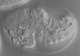 |
Nomarski movie of an embryo demonstrating the Hmr phenotype. Failed epidermal enclosure in the anterior causes extrusion of internal cells. Frames were collected every 2 minutes for 5 hours (8 frames/second display rate). |
Click on the link to load video sequence 2: Video sequence #2 (1.6 Mb)
Return to top
Video sequence 3: Elongation failure in a Hmp embryo
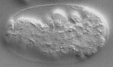 |
Nomarski movie of an embryo demonstrating the Hmp phenotype. Elongation fails and abnormal bulges form on the surface. Frames were collected every 2 minutes for 5 hours (8 frames/second display rate). |
Click on the link to load video sequence 3: Video sequence #3 (1.2 Mb)
Return to top
Video sequence 4: Elongation in a wild-type embryo
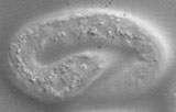 |
Nomarski movie showing development of a wild-type embryo. Frames were collected every 2 minutes for 5 hours (8 frames/second display rate). |
Click on the link to load video sequence 4: Video sequence #4 (1.9 Mb)