


The blastula is a hollow ball of cells organized into an epithelial monolayer. Midway through the blastula stage, the embryo secretes hatching enzyme, which results in proteolytic degradation of the fertilization envelope. The vegetal pole epithelium thickens to form the vegetal plate, which will give rise to primary mesenchyme cells and the archenteron during gastrulation.
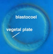
Left: Unhatched blastulae (micrograph courtesy of Dr. David Epel, Hopkins Marine Station). Right: A L. variegatus hatched blastula, showing the thickened vegetal plate and obvious blastocoel (micrograph courtesy of Dr. Chuck Ettensohn, Carnegie-Mellon Univ).
The epithelium is lined on its outer, or apical, surface by two extracellular matrices: (i) an inner apical lamina and (ii) a more exterior hyaline layer. Both are attached to the apices of the cells in the wall of the blastula, which extend microvilli into these extracellular matrix layers.
Fate mapping studies by Hörstadius, Cameron and Davidson; Wray and McClay; and Logan and McClay allow a fairly detailed map of regions of the blastula that will generate the tissue of the gastrula and larva.