Both primary and secondary
mesenchyme cells localize to specific regions of the
embryo, a striking example of pattern
formation. The sea urchin embryo is an excellent
system for examining how cues to which mesenchymal cells
respond are presented within the context of an intact
embryo. In particular, experimental embryology has been
used very successfully to identify cell-cell interactions
in the sea urchin embryo that contribute to mesenchymal
patterning.
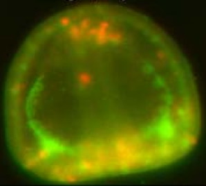
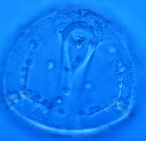
As an aid to such studies, the two types of mesenchyme express different genes, such as cell surface molecules and transcription factors, as the image below shows.
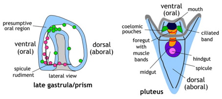
A schematic showing the localization of two types of mesenchyme in the sea urchin embryo. Pink: primary (skeletogenic) mesenchyme; green; secondary mesenchyme. Image by Jeff Hardin, Univ. of Wisconsin.
Both primary and secondary mesenchyme cells have striking localization patterns.