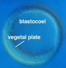
The blastula is a hollow ball of cells organized into an epithelial monolayer (i.e., it is a single cell layer thick). Midway through the blastula stage, the embryo secretes hatching enzyme, a metalloprotease, which results in proteolytic degradation of the fertilization envelope. The vegetal pole epithelium thickens to form the vegetal plate, which will give rise to primary mesenchyme cells and the archenteron during gastrulation. The blastula epithelium is lined on its outer, or apical, surface by two extracellular matrices: (i) an inner apical lamina and (ii) a more exterior hyaline layer. Both are attached to the apices of the cells in the wall of the blastula, which extend microvilli into these extracellular matrix layers.
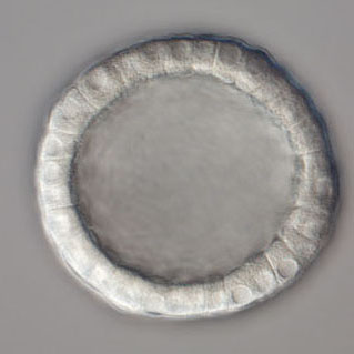
A Dendraster blastula (micrograph courtesy of George von Dassow, Center for Cell Dynamics, Univ. of Washington).
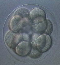
Click on the thumbnails to see movies of cleavage through the early blastula stage in Dendraster, as well as tubulin dynamics in a Dendraster embryo (courtesy of George von Dassow, Center for Cell Dynamics, Univ of Washington). The movie on the right is a time lapse sequence of P. lividus development through hatching, courtesy of Christian Gache and Thierry Lepage, Villefranche-sur-Mer.
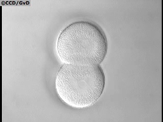
Cleavage in Dendraster (14.5 Mb)
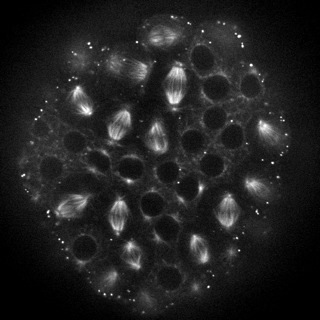
Tubulin in Dendraster (10.8 Mb)
Development through hatching (1.4 Mb)