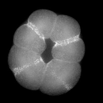
Alteration of cleavage at the 3rd division in L. pictus by pressure. From a movie by Jeff Hardin, Univ. of Wisconsin.
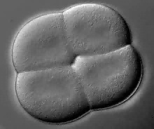
Cleavage pattern can be altered by compressing embryos under a coverslip, forcing all cleavages to occur in the same plane. This technique can be used to address how important specific oriented divisions are for normal development in a variety of species. The movie below, by Jeff Hardin, shows how cleavage is altered at the 3rd division by pressure. In this case, a L. pictus embryo was compressed under a coverslip.
|
|
|
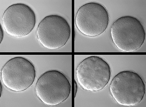
A Dendraster excentricus zygote injected with C3 transferase. Courtesy of George von Dassow, Center for Cell Dynamics, Univ. of Washington.
Rho activity can be monitored using a fluorescent protein that binds to the activated form of Rho. The following two movies (courtesy of Bill Bement, Univ. of Wisconsin-Madison) show that active Rho is recruited to cleavage furrows in echinoderm embryos.
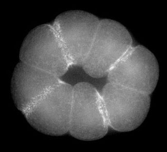
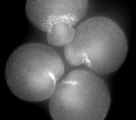
Rho activity monitored using a fluorescent reporter at the 3rd division in S. drobachiensis. From a movie by Bill Bement, Univ. of Wisconsin.
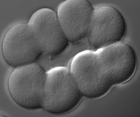
Two movies show how Rho activity dynamically changes during cytokinesis. In the movie on the left, an 8-cell sand dollar embryo has been compressed so that all blastomeres cleave in the same plane. Note how active Rho appears at all cleavage furrows. In the movie on the right, micromeres form at the 4th cleavage in a sea urchin embryo. Active Rho again localizes to cleavage furrows, but the placement of the cleavage furrow is asymmetric along the animal-vegetal axis, resulting in smaller daughter cells (micromeres) and their larger siblings (macromeres).
 |
 |
| Rho activity at 3rd cleavage (1.2 Mb) | Rho activity in micromeres (1.9 Mb) |