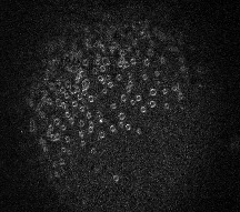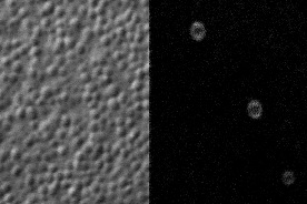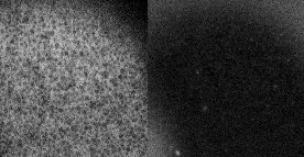
The movies below use fluorescence microscopy to visualize individual cortical granule fusion events (courtesy of Dr. Mark Terasaki, Univ. of Connecticut). The image shows one frame from such a movie.

Cortical granule release
visualized using lipophilic dye (courtesy Mark Terasaki,
Univ. of Connecticut)
As you will see if you play the movie on the right, fusions sweep over the egg surface, beginning with the site of sperm entry, which is evident as the site where calcium elevation occurs first.
 |
 |
 |
|
Cortical granule
release visualized using lipophilic dye (0.7
Mb)
|
Cortical granule
release at high magnification (0.5 Mb)
|
Calcium elevation and
cortical granule release (0.6 Mb)
|