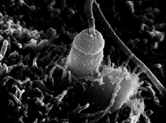
Scanning elecron micrograph of a sea urchin sperm penetrating an egg. The fertilization cone is visible to the right of the sperm head. Image courtesy of Gerald Schatten, Univ. of Pittsburgh.
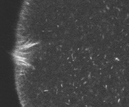
Fertilization cone visualization in Lytechinus. Actin has been visualized using rhodmaine phalloidin. Courtesy of Mark Terasaki, Univ. of Connecticut.
Click on the thumbnail to see a movie of fertilization cone formation, courtesy of Mark Terasaki, Univ. of Connecticut. |
|
fertilzation cone
visualized via phalloidin (1.2 Mb)
|
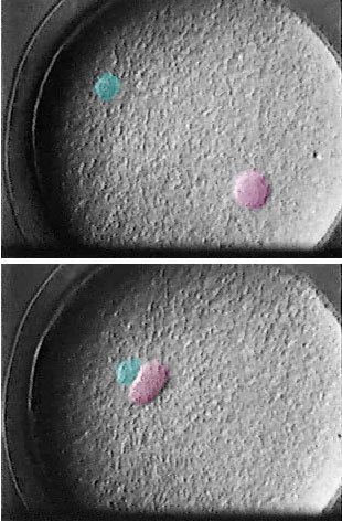
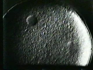
Pronuclear migration in L. variegatus. The male pronucleus is colorized in blue; female in pink. Frames from a movie courtesy of Shinya Inoue, Marine Biological Laboratory, Woods Hole.
Pronuclear migration is a dramatic process in many species, including sea urchins. Microtubules, nucleated by the sperm's centrosome, serve as guides for the movement of the female pronucleus, a process mediated by the microtubule motor cytoplasmic dynein.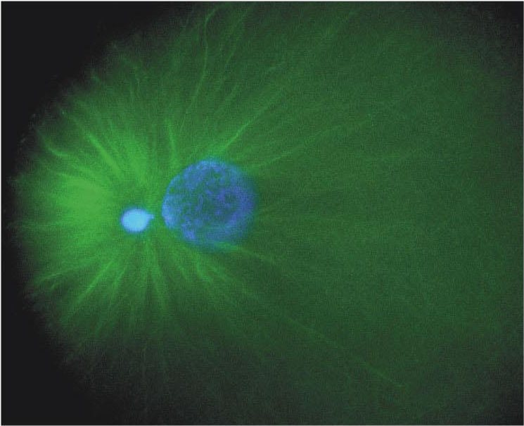
Microtubules during pronuclear migration in L. pictus. The small male pronlucleus and the larger female pronucleus are stained blue with a DNA dye; microtubules are immunostained in green. Courtesy of Jon Holy, Univ. of Minnesota-Duluth.
In the classic footage below, courtesy of Shinya Inoue, Marine Biological Laboratory, Woods Hole, the migration of the female pronucleus towards the male pronucleus can be seen. Because it is a microtubule-dependent process, pronuclear migration can be reversibly blocked by microtubule depolymerizing drugs, such as colcemid. When the drug is washed out, microtubules can reform, and migration can resume.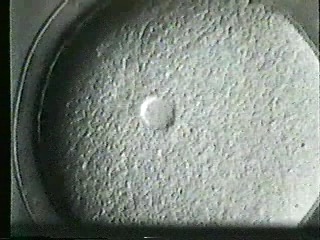 |
 |
|
Pronuclear
migration in L. variegatus (5.7 Mb)
|
Migration
reversibly blocked by colcemid (5.7 Mb)
|
Imaging of pronuclear migration can also be performed by labeling membranous structures within the egg with a dye, as the image below from Mark Terasaki, Univ. of Connecticut, shows.
Imaging of pronuclear migration in Lytechinus. The egg's contents have been labeled using a liophilic dye (DiI). Courtesy of Mark Terasaki, Univ. of Connecticut.
Click on the thumnail to see the corresponding movie...
|
pronuclear
migration imaged using DiI (0.3 Mb)
|