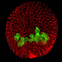
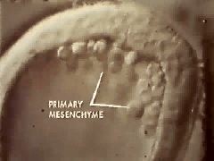
A L. variegatus mesenchyme blastula, viewed from the side, showing ingressing primary, or skeletogenic, mesenchyme cells (PMCs). Image courtesy of David McClay, Duke Univ.
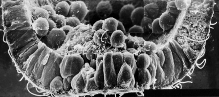
A L. variegatus
mesenchyme blastula processed
for scanning electron microscopy and fractured through the
vegetal plate to show the process of ingression (photograph
courtesy of Dr. John Morrill, Univ. of South
Florida).
 |
|
 |
|
Classic footage of
ingression in Dendraster excentricus.
Size: 1.1 Mb.
|
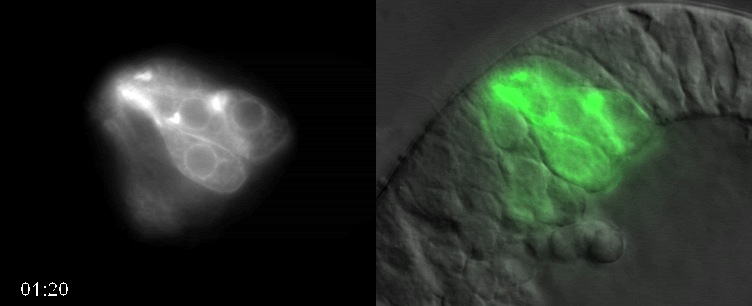
Movie of PMC
ingression in Lytechinus variegatus. Size:
4.7 Mb
|
Movie of PMC
ingression in Lytechinus variegatus (PMCs
express the green fluorescent protein, GFP).
Size: 7.1 Mb
|
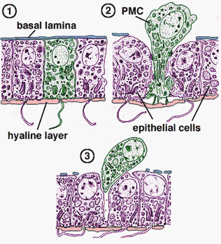
Although the mechanisms of ingression are not fully understood, several events are known to accompany ingression. The best characterized changes in PMCs involve changes in their adhesive properties. PMCs must undergo several changes in their adhesive properties in order to detach from the epithelium of the vegetal plate and ingress into the interior: (1) they must lose affinity for neighboring epithelial cells that remain in the vegetal plate; (2) they must lose affinity for the hyaline layer on the exterior of the embryo; and (3) they presumably must gain affinity for the basal lamina, since PMCs later migrate within the blastocoel. Hideki Katow and Michael Solursh used transmission electron microscopy to show that PMCs undergo a dramatic reorganization of their cytoskeleton and their contacts with these three regions during ingression. Rachel Fink and David McClay used cell adhesion assays to show that PMCs undergo quantitative changes in their adhesive affinities.
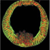
Immunostaining for cadherin (green) and b- catenin (red) in a Lytechinus variegatus early mesenchyme blastula (left) and a later mesenchyme blastula. Note loss of b-catenin in PMCs that have ingressed. Micrograph courtesy of David McClay, Duke Univ.