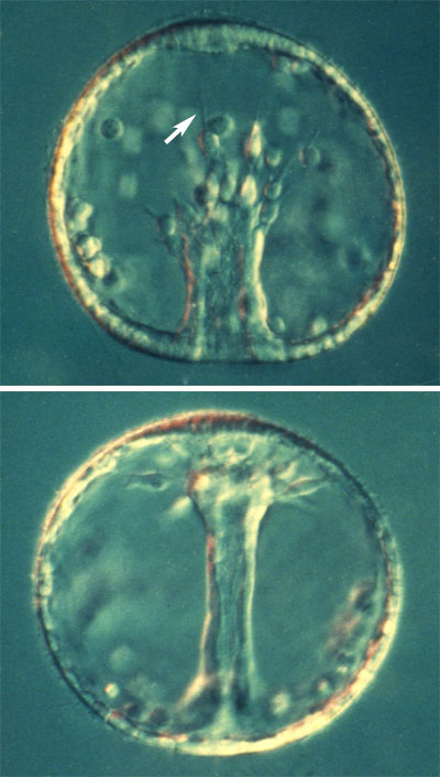
L. pictus midgastrula (top) and late gastrula (bottom). Note the filopodia extended by secondary mesenchyme cells (SMCs; arrow). Images by Jeff Hardin, Univ. of Wisconsin.
Secondary invagination involves the elongation of the archenteron across the blastocoel, where it attaches to the ectoderm near the animal pole of the embryo. The onset of secondary invagination correlates with the appearance of long, thin filopodia extended by secondary mesenchyme cells (SMCs) at the tip of the archenteron. Under a variety of circumstances, there is substantial evidence that SMCs are important for extension of the archenteron during secondary invagination. However, experiments done in the 1980s by Charles Ettensohn and myself suggest that SMCs are not the sole driving force for archenteron elongation. To see a movie overview of archetneron elongation in L. variegatus, courtesy of Charles Ettensohn, click on the lefthand thumbnail below. To see a classic piece of film footage of archenteron elongation in Dendraster by Richard Cloney, click on the righthand thumbnail below.
 |
 |
|
|
|
The next two pages discuss our current view of archenteron elongation.