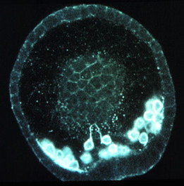
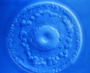
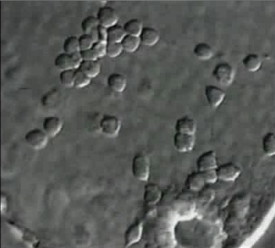
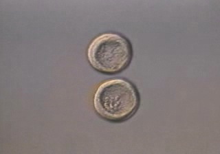
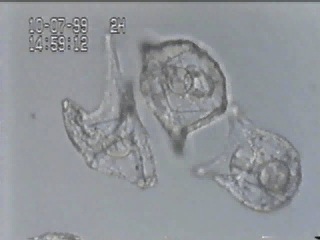
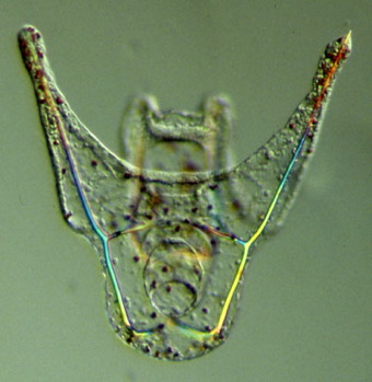
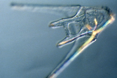
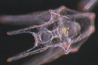
Skeletogenic (primary) mesenchyme cells (PMCs) migrate within the blastocoel, extending filopodia as they arrange themselves into a precise pattern. In the confocal image at the left, two PMCs are extending filopodia towards the vegetal pole. PMCs initially aggregate into two ventrolateral clusters as a prelude to forming the bona fide skeleton. In the image at left, the clusters are beginning to form. As PMCs aggregate, they begin to secrete small crystalline skeletal elements. In the beautiful image below, a small "propeller", which is the beginning of a spicule, is visible.
Lytechinus
variegatus late
gastrula, ventral view. Image courtesy of Charles
Ettensohn, Carnegie-Mellon University.
 |
|
|
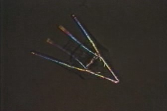
Eventually, the PMCs secrete a calcium carbonate-containing skeleton that gives the sea urchin larva - the pluteus - its characteristic shape ["Pluteus" comes from a Greek word for "easel"; if you turn the pictures below upside down, you can see why early embryologists gave it this name.] Each of the skeletal rods is called a spicule, and the pattern of these spicules is very precise. Click on the thumbnails to see movies of swimming gastrulae and larvae visualized using polarization optics, which allows spicules to be visualized easily (Left and middle: courtesy of Christian Gache and Thierry Lepage, Villefranche-sur-Mer; right courtesy of Rachel Fink, Mt. Holyoke College)
 |
 |
 |
|
Spicules in P.
lividus late gastrulae (0.8 Mb)
|
Spicules in P.
lividus plutei (1.3 Mb)
|
Spicules in
Lytechinus plutei (3.9 Mb)
|
The images of the dorsal and ventral surfaces of a L. variegatus pluteus below show just how precise the skeletal pattern is..
Top: A L. variegatus
pluteus viewed from the
dorsal (aboral) side. Bottom: A L. variegatus
pluteus viewed from the
ventral (oral) side. Images by Jeff Hardin, Univ, of Wisconsin.
 |
|
Feeding P.
lividus pluteus (0.4 Mb)
|