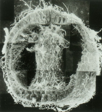
A L. variegatus late gastrula, processed via scanning
electron microscopy (SEM) and fractured open to reveal the
archenteron making contact with the animal pole. Image
courtesy of John Morrill, Univ. of South Florida.
Secondary mesenchyme cells
(SMCs) reliably attach the tip of the archenteron to a
region that will become the mouth of the later larva. This
oral (ventral) attachment site presumably expresses
specific adhesion and.or guidance molecules to which SMCs
respond. SMCs that make contact with this region change
their behavior dramatically, making extremely long-lived
filopodial connections to this site, as this classic
footage from my days as a postdoctoral fellow shows.
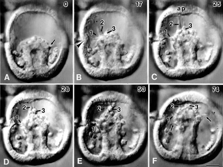
Secondary mesenchyme cells
make stable contacts with a specific region near the animal
pole. Frames from a time-lapse sequence of gastrulation
in L.
variegatus. Time
is shown in minutes in the upper right. Three filopodia are
followed during the course of the movie. Note that
filopodium #3 remains connected where it makes its
attachment for more than 45 minutes. Images by
Jeff Hardin, Univ. of Wisconsin.