 THE HARDIN LAB
C. elegans Enabled Exhibits Novel Interactions with N-WASP, Abl, and Cell-Cell Junctions
Mark Sheffield1, Timothy Loveless2, Jeff Hardin1,2,3 and Jonathan Pettitt4
1Program in Genetics, University of Wisconsin, 1117 West Johnson Street, Madison, Wisconsin 53706 2Cellular and Molecular Biology Graduate Program, University of Wisconsin, 1117 West Johnson Street, Madison, Wisconsin 53706 3Department of Zoology, University of Wisconsin, 1117 West Johnson Street, Madison, Wisconsin 53706 4School of Medical Sciences, University of Aberdeen Institute of Medical Sciences, Aberdeen AB25 2ZD, United Kingdom
Supplemental material for the following paper:
Sheffield, M.S., Loveless, T., Hardin, J., and Pettitt, J. (2007). C. elegans Enabled exhibits novel interactions with N-WASP, Abl, and cell-cell junctions. Curr. Biol. 17, 1791-1796. PubMed
Summary
Video sequence 1
Video sequence 2
Video sequence 3
Video sequence 4
Video sequence 5
Summary
Ena/VASP proteins are associated with cell-cell junctions in cultured mammalian cells and Drosophila epithelia, but they have only been extensively studied at the leading edges of migratory fibroblasts, where they modulate the protrusion of the leading edge. They act by regulating actin-filament geometry, antagonizing the effects of actin-capping protein. Embryos lacking the C. elegans Ena/VASP, UNC-34, display subtle defects in the leading edges of migrating epidermal cells but undergo normal epidermal morphogenesis. In contrast, embryos lacking both UNC-34 and the C. elegans N-WASP homolog have severe defects in epidermal morphogenesis, suggesting that they have parallel roles in coordinating cell behavior. GFP-tagged UNC-34 localizes to the leading edges of migrating epidermal cells, becoming redistributed to new junctions that form during epidermal-sheet sealing. Consistent with this, UNC-34 contributes to the formation of cadherin-based junctions. The junctional localization of UNC-34 is independent of proteins involved in Ena/VASP localization in other experimental systems; instead, junctional distribution depends upon the junctional protein AJM-1. We also show that Abelson tyrosine kinase, a major regulator of Enabled in Drosophila, is not required for UNC-34/Ena function in epithelia. Instead, our data suggest that Abelson kinase acts in parallel to UNC-34/Ena, antagonizing its function.
Return to top
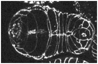
Movie S1 (1628K). Ventral Enclosure in the Wild-Type Visualized with AJM-1::GFP. A projection of from a 4D multiphoton movie of an embryo expressing ajm-1::gfp (ventral view, anterior is to the left). The interval between frames is 180 s.
Download

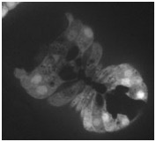
Movie S2 (2377 K). Morphogenesis Defects in an unc-34(gm104);wsp-1(RNAi) Embryo Visualized with AJM-1::GFP. Dorsal (right) and ventral (left) projected views of the same unc-34(gm104);wsp-1(RNAi) embryo from a 4D multiphoton movie of an embryo expressing ajm-1::gfp (anterior is to the left in each case). The interval between frames is 180 s.
Download
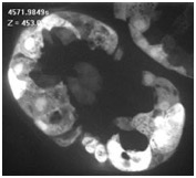
Movie S3 (1908 K). Leading Cell Protrusive Zone Activity in the Wild-Type Visualized with a dlg-1 Transcriptional Reporter. Projections of dlg-1Δ7::gfp expression from a spinning-disc confocal 4D movie (ventral view, anterior is to the upper left). The interval between frames is 50 s.
Download
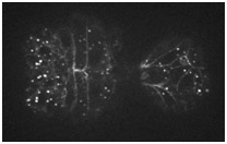
Movie S4 (3136 K). LCPZ activity in an unc-34(gm104);wsp-1(RNAi) Embryo Visualized with a dlg-1 Transcriptional Reporter. Projections of dlg-1Δ7::gfp expression from a spinning-disc confocal 4D movie (ventral view, anterior is to the upper left). The interval between frames is 50 s.
Download
Movie S5 (4372 K). UNC-34::GFP Localization in the Wild-Type during Ventral Enclosure. Projections from a spinning-disc confocal 4D movie (ventral view, anterior is to the left). The interval between frames is 20 s.
Download
Return to top