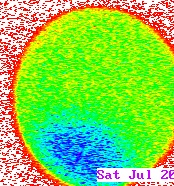
Pseudocolor image from a movie courtesy of Dr. Gerald Schatten (Univ. of Pittsburgh). High levels of calcium are shown as blue; low levels as red. See if you can tell where the sperm entered the egg!
Calcium dynamics can be quantitatively measured using indicators, and depicted as a "4-dimensional" movie. This image, courtesy of Michael Whitaker (Univ. of Newcastle) depicts elevated calcium levels using both color and a third axis, in which greater height also indicates higher calcium ion concentration. Again, see if you can tell where the sperm entered the egg!
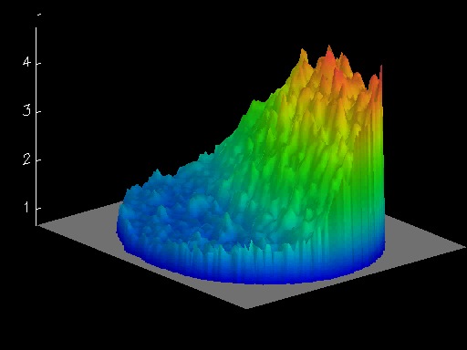
4-dimensional depiction of calcium levels in a L. pictus zygote. From a movie courtesy of Michael Whitaker, Univ. of Newcastle.
Calcium dynamics can be imaged nicely in movies. Click on the corresponding thumnails to see movies corresponding to the two images shown above.
|
|
|
|
|
|
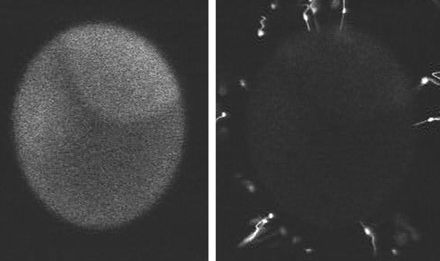
Two additional movies (provided by Dr. c), show that calcium elevation iniates near the site of sperm entry, and that calcium elevation correlates with where fertilization envelope formation begins. In the first movie, fluorescently labeled sperm are used to fertilize an egg which has beeninjected with a caclium indicator. Notice that in this movie the calcium levels elevate in the same location as the first place where the fertilization envelope lifts off of the egg surface.
|
|
 |
|
|
|