Sea urchin sperm can be obtained in large quantities,
because sea urchins, like many marine invertebrates,
engage in broadcast spawning, i.e., they release
large quantities of gametes into the sea water. Undiluted
semen contains a large quantity of sperm - as many as
1010 - 1011 cells/ml!
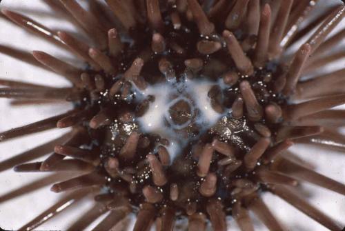
A male Arbacia punctulata spawning its whitish semen. Image courtesy of Thomas Gensch, Forschungszentrum Jülich.
Sperm swim by means of a prominent flagellum, composed of a core of microtubules, whose sliding is powered by flagellar dynein. This array of microtubules and associated motor and linker proteins is known as an axoneme. The midpiece of the sperm contains a prominent array of mitochondria, which are required to produce huge amounts of ATP, whose hydrolysis powers the conformational changes in flagellar dynein that mediate microtubule sliding.
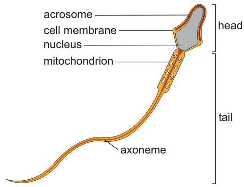
Image courtesy of Thomas Gensch, Forschungszentrum Jülich.
Most flagella, like cilia, have a characteristic “9+2” structure, i.e., two central singlet microtubules are encircled by nine outer doublet microtubules. The outer and inner dynein arms slide along each outer doublet microtubule. The following diagram shows how the inner and outer doublet microtubules of the axoneme are connected to dynein.
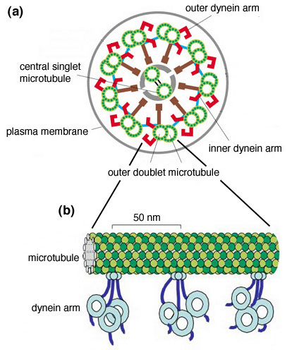
Internal structure of the axoneme. Adapted from an image courtesy of Takashi Ishikawa, Swiss Federal Institute of Technology, Zurich.
Sea urchin sperm move their tails using a helical motion, as shown here in this animation.
Animation courtesy of Sea Urchin Embryologysite.
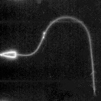
Finally, as the movie below shows, flagellar bending and the sliding of microtubules can be examined by analyzing the movements of small beads placed along the surface of a flagellum treated with detergent. The beads move apart as the wave of flagellar bending passes by, indicating that flagellar bending is mediated by microtubule sliding.
Description: This sea urchin spermatozoon was treated with detergent to remove the cell membrane, and then transferred to a solution containing a relatively low concentration of MgATP, to reactivate the bending of its flagellum. Before reactivation, the spermatozoa were exposed to a suspension of 40 nm gold beads. Some of these beads attach to the outer doublet microtubules that comprise the flagellar axoneme. In this case, two beads were attached to doublet microtubules on opposite sides of the flagellum. As the flagellum bends, the sliding between these doublet microtubules is demonstrated by the movement of the two beads. Movie courtesy of Charles Brokaw, California Institute of Technology.
A male Arbacia punctulata spawning its whitish semen. Image courtesy of Thomas Gensch, Forschungszentrum Jülich.
Sperm swim by means of a prominent flagellum, composed of a core of microtubules, whose sliding is powered by flagellar dynein. This array of microtubules and associated motor and linker proteins is known as an axoneme. The midpiece of the sperm contains a prominent array of mitochondria, which are required to produce huge amounts of ATP, whose hydrolysis powers the conformational changes in flagellar dynein that mediate microtubule sliding.
Image courtesy of Thomas Gensch, Forschungszentrum Jülich.
Most flagella, like cilia, have a characteristic “9+2” structure, i.e., two central singlet microtubules are encircled by nine outer doublet microtubules. The outer and inner dynein arms slide along each outer doublet microtubule. The following diagram shows how the inner and outer doublet microtubules of the axoneme are connected to dynein.
Internal structure of the axoneme. Adapted from an image courtesy of Takashi Ishikawa, Swiss Federal Institute of Technology, Zurich.
Sea urchin sperm move their tails using a helical motion, as shown here in this animation.
Animation courtesy of Sea Urchin Embryologysite.
Finally, as the movie below shows, flagellar bending and the sliding of microtubules can be examined by analyzing the movements of small beads placed along the surface of a flagellum treated with detergent. The beads move apart as the wave of flagellar bending passes by, indicating that flagellar bending is mediated by microtubule sliding.
Description: This sea urchin spermatozoon was treated with detergent to remove the cell membrane, and then transferred to a solution containing a relatively low concentration of MgATP, to reactivate the bending of its flagellum. Before reactivation, the spermatozoa were exposed to a suspension of 40 nm gold beads. Some of these beads attach to the outer doublet microtubules that comprise the flagellar axoneme. In this case, two beads were attached to doublet microtubules on opposite sides of the flagellum. As the flagellum bends, the sliding between these doublet microtubules is demonstrated by the movement of the two beads. Movie courtesy of Charles Brokaw, California Institute of Technology.
Sliding of microtubules in sperm (0.2 Mb)