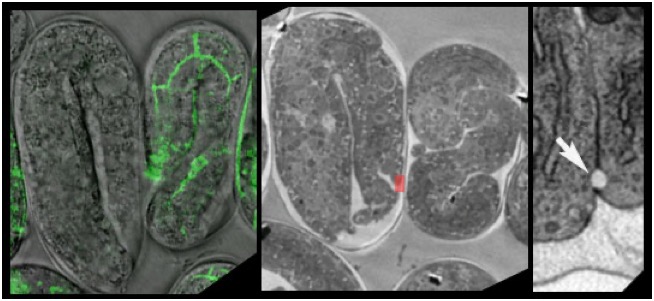
(Left) Two elongating C. elegans embryos mutant for ajm-1, which encodes a junctional protein imaged in an agar mount using transmitted light (gray) and confocal microscopy. One expresses AJM-1::GFP (green), and so it is rescued. After processing for TEM (middle), ultrastructural details can be examined. (Right) The small red rectangle in the middle panel at higher magnification, showing a junctional "bubble" (arrow) [Paul Sims].
We developed techniques to perform dynamic 4d imaging of wild-type and mutant embryos followed by high-resolution imaging of ultrastructure using high-pressure freezing and immunoEM. Our goal was to bridge the gap between dynamic analysis of living embryos at the level of light microscopy and the fine structure of cell-cell junctions during morphogenesis.